

AI-Cofolding
Predict a complex's 3D structure and binding affinity with Boltz-2Give protein sequences and, optionally, a ligand, DNA or RNA, and Boltz-2 predicts how they fold and fit together. For a small-molecule ligand it can also predict how strongly it binds, so you can check a pose before spending lab time.
Boltz-2 (boltz 2.2.1) · Doc updated Oct 14, 2026
- Input
- Sequences, structures, SMILES
- Output
- 3D complex, confidence, affinity
- Engine
- Boltz-2
- Cost
- From 100 cr, quoted before submit
- Standard preset
- About 45 min
When to use
- Your target has no experimental structure, or not with your ligand bound
- You want to see how a hit from Virtual Screening sits in the pocket before ordering it
- You need a quick binding estimate for a handful of ligands against one protein
- You want to model a protein with a partner chain, DNA or RNA
A predicted structure is a model, not a measurement. Treat low-confidence regions with care, and use the affinity value to rank ligands rather than as a lab-grade number.
How it works
- 01Read the inputs
Each sequence, structure chain or SMILES becomes one chain of the complex, lettered A, B, C in the order you add them.
- 02Fold together
Boltz-2 predicts all chains at once, so the protein can shape itself around the ligand. Steering potentials keep the ligand's chemistry, including its stereocenters, physically valid.
- 03Sample and rank
It draws many candidate structures (diffusion samples) and ranks them by confidence. Model 0 is the best.
- 04Score binding
If you turned affinity on, a separate head predicts how strongly the ligand binds and how likely it is to bind at all.
Input formats
| Item | Accepted | Notes |
|---|---|---|
| Protein, DNA, RNA | Pasted sequence (FASTA headers are fine), 4-character PDB ID, or a .pdb/.cif upload | From a structure you pick the chain; if you don't, the longest chain is used. At least one protein is required |
| Ligand | SMILES, or an SDF upload | SDF: up to 50 molecules and 10 MB. Salts and counter-ions are removed and the largest fragment is kept |
| MSA (optional, protein) | A precomputed .a3m file, up to 50 MB | Without one, the chain folds from its sequence alone (single-sequence mode) |
| Template (optional, protein) | .cif or .pdb | Guides the fold toward a known structure |
| Size | Up to 10 components and 1,437 residues in total | Larger inputs are rejected before you are charged |
Tutorial: EGFR with erlotinib
PDB 1M17 is the kinase domain of EGFR, a lung cancer target, with the drug erlotinib bound (ligand code AQ4). Here we ask Boltz-2 to rebuild that complex from the sequence and the SMILES alone, and check whether erlotinib lands in the same pocket and pose as in the crystal. Run example at the top of this page fills all of this in for you.
Add the protein
In Components, add a protein and enter the PDB ID
1M17. Pick chain A. The preview pane shows the structure.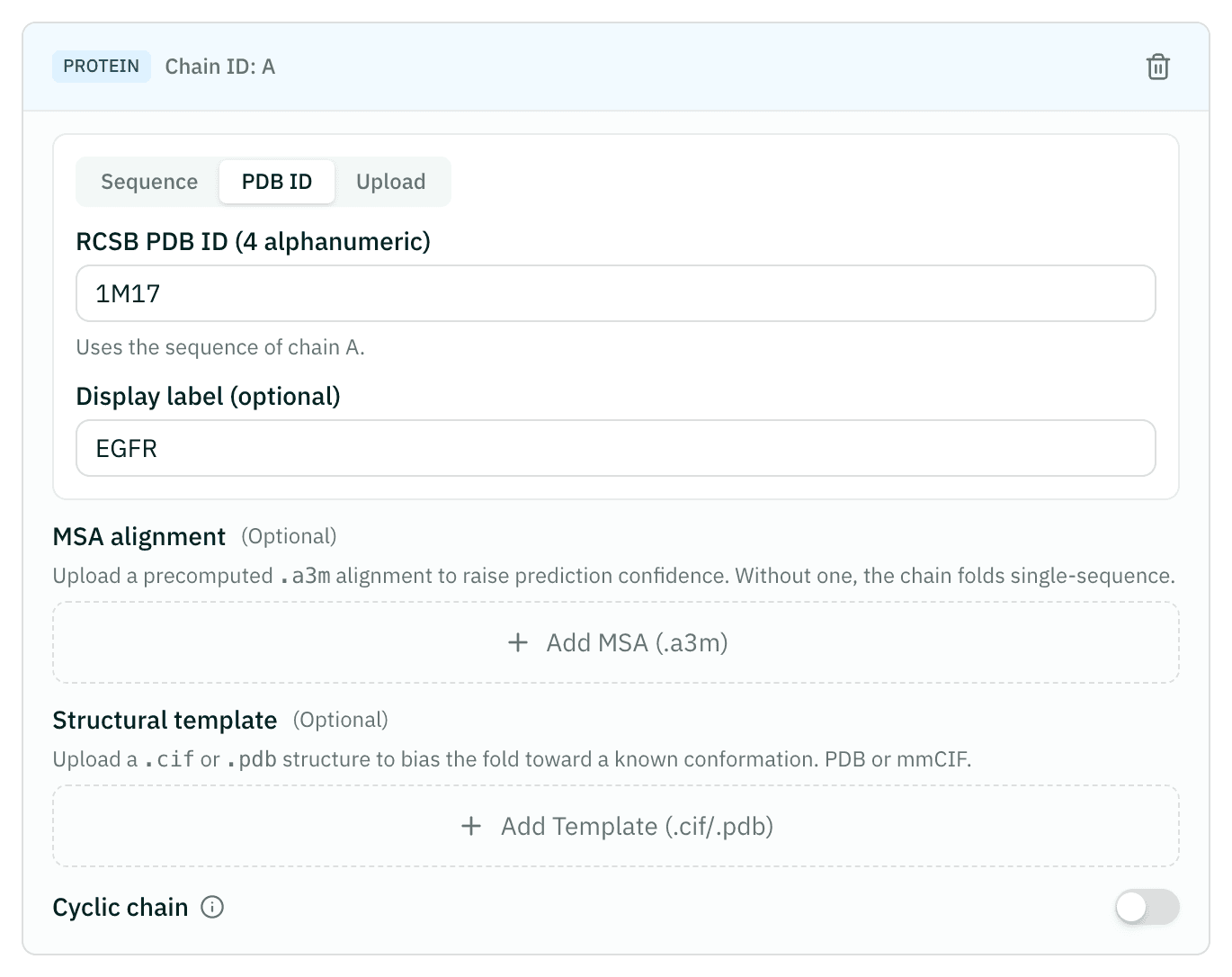
Add erlotinib
Add a ligand and paste its SMILES:
COCCOc1cc2ncnc(Nc3cccc(C#C)c3)c2cc1OCCOC. Leave Calculate binding affinity off for this example: the pose is the question here, and affinity adds 35–60 minutes. Turn it on when you want a binding estimate (one ligand per job).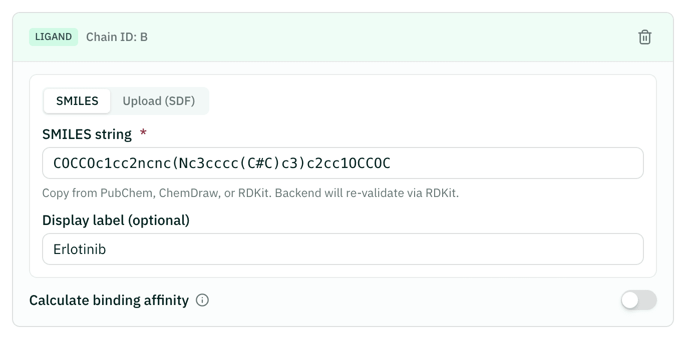
Choose a preset
In Advanced Settings, choose Quick (5 recycling steps, 20 samples), enough for a first look. Standard and High Accuracy take longer for a better model.
Check the quote and submit
In Project Settings, name the project. The summary shows the quoted credits and time; submit when they look right. We email you when the job finishes.
Read the result
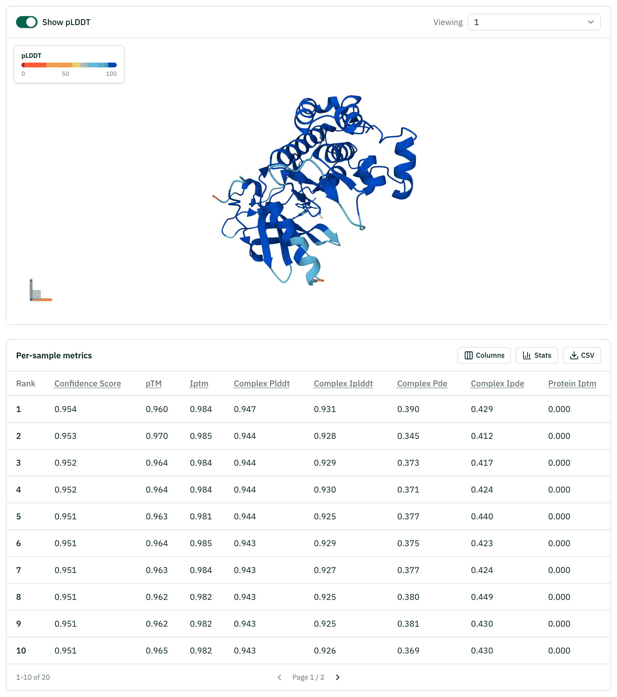
Open the project. Check ipTM and pLDDT for model 0, color the structure by pLDDT, and see whether erlotinib sits in the ATP pocket the way it does in 1M17.
Reading the results
| Value | Meaning | How to read it |
|---|---|---|
| Confidence score | Boltz-2's overall ranking score (0–1) | Higher is better; samples are ranked by it |
| ipTM | Confidence in how the chains sit relative to each other (0–1) | The key number for a complex. Below 0.5, doubt the binding pose |
| pTM | Confidence in the overall fold (0–1) | Higher is better |
| pLDDT | Per-residue confidence (0–1 in the table) | Below 0.6 means a shaky model. The 3D view colors it with the usual 50/70/90 bands |
| PDE, iPDE | Predicted distance error over the complex and at the interface | Lower is better |
| PAE heatmap | Predicted position error between every pair of residues, in Å | Dark blocks between two chains mean their relative placement is trusted |
| pIC50 | Predicted affinity, 6 minus Boltz-2's log10 IC50 (µM) | Higher means tighter binding. Each step of 1 is a tenfold change |
| Binding probability | Chance the ligand binds at all (0–1) | Use it to separate binders from non-binders before comparing pIC50 |
When model 0 has pLDDT below 0.6 or ipTM below 0.5, the result page shows tips for improving the run, such as adding an MSA.
Example result: EGFR 1M17 with erlotinib
A real job, shown read-only. Sign in to run the same setup yourself.
| Model | Confidence | ipTM | pLDDT | pTM |
|---|---|---|---|---|
| model_0 | 0.954 | 0.984 | 0.947 | 0.960 |
| model_1 | 0.953 | 0.985 | 0.944 | 0.970 |
| model_2 | 0.952 | 0.984 | 0.944 | 0.964 |
| model_3 | 0.952 | 0.984 | 0.944 | 0.964 |
| model_4 | 0.951 | 0.981 | 0.944 | 0.963 |
Cost and time
The price follows the GPU time your inputs need: protein length, number of samples, recycling steps, and whether you add an MSA or affinity. The wizard asks the server for a quote as you edit, and you pay exactly that quote at submit. The minimum is 100 cr (1 cr is $0.01).
| Preset | Recycling, samples | Wizard's rough guide |
|---|---|---|
| Quick | 5, 20 | About 15 min |
| Standard | 10, 50 | About 45 min |
| High Accuracy | 20, 100 | About 2 hours |
Affinity adds roughly 35–60 minutes; the example leaves it off. The example above took about 27 minutes from submit to results and cost 188 cr.
If the engine fails, the credits come back in full. If you stop a job yourself, half comes back. A job is stopped after 5 hours.
Citations
If you use these results in a report or paper, please cite the methods below.
- Passaro S, Corso G, Wohlwend J, et al.. Boltz-2: Towards accurate and efficient binding affinity prediction. bioRxiv (2025). doi:10.1101/2025.06.14.659707Structure and affinity prediction
- Wohlwend J, Corso G, Passaro S, et al.. Boltz-1: Democratizing biomolecular interaction modeling. bioRxiv (2024). doi:10.1101/2024.11.19.624167Boltz structure model
- Stamos J, Sliwkowski MX, Eigenbrot C. Structure of the epidermal growth factor receptor kinase domain alone and in complex with a 4-anilinoquinazoline inhibitor. J Biol Chem 277, 46265-46272 (2002). doi:10.1074/jbc.M207135200EGFR–erlotinib structure used in the tutorial (PDB 1M17)